Introduction
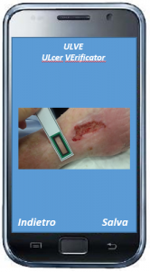
ULVE is a program for the analysis of pressure, diabetic and vascular ulcers, which permits to make an objective assessment of the state of the injury simply using a smartphone and a reference marker to be placed near the area of interest.

The application, therefore, allows the doctor to remotely monitor the evolution of ulcers without the patient having to go to the clinic.
Functionalities
The functionalities supported by the application are the following:
- Acquisition of the photo (manual);
- Segmentation of the lesion (automatic);
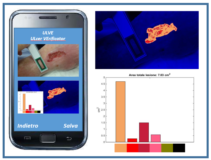
- Area calculation (automatic);
- Tissue analysis (automatic);
Why to use ULVE
- It is a non-invasive tool;
- It is an objective evaluation tool;
- It is easy to use.
Advantages and benefits
ULVE is a non-invasive tool for the patient and its use allows the doctor to have objective and quantitative feedback on the state of the injury. The image acquisition process is simple and low-cost.
Furthermore the application:
- Supports the process of treatment of ulcers;
- Improves the quality of care;
- Helps to recognize and quantize the tissues constituting the ulcer;
- Improves the monitoring of the healing process;
- Automates clinical documentation;
- Improves communication and collaboration for method sharing;
- Reduces the days of hospitalization;
- Promotes home care;
- Promotes telemedicine;
Guide to use
- Image Acquisition
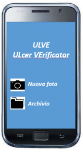
As the application turns on, the user can choose to consult the archive of the saved files or proceed with the acquisition of a new picture.
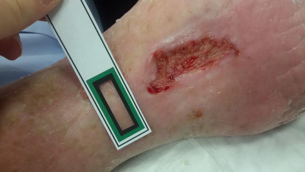
The photo of the area where the ulcer is present is taken by positioning the reference marker near the lesion (at a distance of about 1 cm). The ulcer and the green portion of the marker must be completely framed in the image.
- Analysis
Once the image acquisition is done, the SW automatically generates the mask that identifies the area to be analyzed.
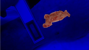
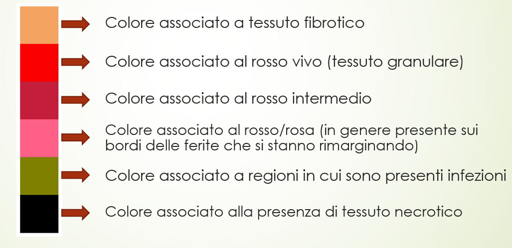
The SW then performs the colorimetric analysis of the tissues belonging to the mask previously identified. In particular, it associates each pixel of the image with one of the 6 defined colorimetric classes, representative of the tissues that may be present in this type of ulcers; it also returns a histogram to quantify the colors present in the lesion.